Sunday, June 30, 2019

About kidneys – The hero of your body.


This section of our site has a lot of general information about kidneys, what can go wrong with them, ways to look after your kidneys and information about a kidney health check.

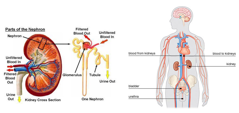
The kidneys are two bean shaped organs found in the middle of your abdomen towards the back. (See below picture) They have the very important job of filtering waste and excess fluid out of your blood to make urine. 

The parts of the kidney that filter the blood are called nephrons (from the Greek word nephros meaning kidney). There are about 1 million nephrons in each kidney working all day, every day like little filters “cleaning” your blood.


In addition to their “waste management” role, the kidneys also have the important job of controlling your blood pressure, helping red blood cell production, keeping your blood’s acid/base balance right and contributing to healthy bones.


What can go wrong with your kidneys?


Kidney disease is often referred to as a “silent disease”. Some people can lose 90% of kidney function before feeling sick. Kidney damage can either be short term (Acute Kidney Injury) or long term (Chronic Kidney Disease). Many illnesses can lead to this. A common cause of Acute Kidney Injury is dehydration. Common causes of Chronic Kidney Disease are diabetes, high blood pressure, and inflammation of the nephrons (glomerulonephritis). If the kidney function drops, waste products can build up in your body, high blood pressure is common, anaemia (not enough red blood cells) and bone problems occur.


How to look after your kidneys? 


Maintain a healthy weight. 
Exercise regularly. 
Stop smoking, or don’t start smoking! 
Limit alcohol consumption. 
Drink water to avoid dehydration. 
Get your blood pressure checked. 
Ask your GP if you need a Kidney Health Check 


If you have high blood pressure, diabetes, obesity, heart disease, have had a stroke, have a family history of kidney disease, are a smoker, are aged over 60 years, have had an episode of Acute Kidney Injury, or are Aboriginal or Torres Strait Islander, you are at greater risk of Chronic Kidney Disease. If this applies to you, see your GP and consider a Kidney Health Check.


What is a Kidney Health Check?


A Kidney Health Check combines three tests your GP can do to check (screen) for kidney disease. These include having your blood pressure checked, having a blood test measuring your Estimated Glomerular Filtration Rate, also called eGFR, and a urine test for your Albumin:Creatinine Ratio (see Tests and Procedures). Together, these three tests indicate how well your kidneys are working.
Kidney Diagram - Richard Baer Kidney Specialist Brisbane

“An investment in knowledge always pays the best interest”

– Benjamin Franklin

ALL ABOUT CIRCULATION ( PULMONARY & SYSTEMIC)


The circulatory system is a vast network of organs and vessels that is responsible for the flow of blood, nutrients, hormones, oxygen and other gases to and from cells. Without the circulatory system, the body would not be able to fight disease or maintain a stable internal environment — such as proper temperature and pH — known as homeostasis.


Description of the circulatory system


While many view the circulatory system, also known as the cardiovascular system, as simply a highway for blood, it is made up of three independent systems that work together: the heart (cardiovascular); lungs (pulmonary); and arteries, veins, coronary and portal vessels (systemic), according to the U.S National Library of Medicine (NLM).

In the average human, about 2,000 gallons (7,572 liters) of blood travel daily through about 60,000 miles (96,560 kilometers) of blood vessels, according to the Arkansas Heart Hospital. An average adult has 5 to 6 quarts (4.7 to 5.6 liters) of blood, which is made up of plasma, red blood cells, white blood cells and platelets. In addition to blood, the circulatory system moves lymph, which is a clear fluid that helps rid the body of unwanted material.

The heart, blood, and blood vessels make up the cardiovascular component of the circulatory system. It includes the pulmonary circulation, a "loop" through the lungs where blood is oxygenated. It also incorporates the systemic circulation, which runs through the rest of the body to provide oxygenated blood, according to NLM.

The pulmonary circulatory system sends oxygen-depleted blood away from the heart through the pulmonary artery to the lungs and returns oxygenated blood to the heart through the pulmonary veins, according to the Mayo Clinic

Oxygen-deprived blood enters the right atrium of the heart and flows through the tricuspid valve (right atrioventricular valve) into the right ventricle. From there it is pumped through the pulmonary semilunar valve into the pulmonary artery on its way to the lungs. When it gets to the lungs, carbon dioxide is released from the blood and oxygen is absorbed. The pulmonary vein sends the oxygen-rich blood back to the heart, according to NLM.

The systemic circulation is the portion of the circulatory system is the network of veins, arteries and blood vessels that transports blood from the heart, services the body's cells and then re-enters the heart, the Mayo Clinic noted.


The Pulmonary Circulation


The pulmonary circulation consists of the pulmonary trunk, which extends from the right ventricle and divides into right andleft pulmonary arteries. The right pulmonary artery is wider and longer than the left. Each artery enters the hilum of the lung, where it further divides into smaller branches.
The left pulmonary artery is attached to the aorta through the ligamentum arteriosum, a fibrous remain of ductus arteriosus. If the ductus arteriosus remains patent after birth, this condition is known as patent ductus arteriosus (PDA). In this case, the deoxygenated blood bypasses the lung and enters directly into the aorta. Babies with PDA are cyanosed and need an urgent medical or surgical intervention.
Pulmonary veins arise from each lobe of the lung. The veins from the right upper and middle lobe unite, and a total of four pulmonary veins enter the left atrium.

The Systemic Circulation


The systemic blood vessels present in the thorax include: aortabrachiocephalic trunkbrachiocephalic veinssuperior and inferior vena cavaazygous vein and the vertebral veins. These are discussed individually, below.
  • The aorta is the largest artery in the human body. It is divided into three major parts: the ascending aorta, the arch of aorta, and the descending aorta. The part of the descending aorta within the thorax is known as thoracic aorta. The ascending aorta gives off right and left coronary arteries. It ascends up to the level of sternal angle.
    • The arch of aorta has three branches:
      • Brachiocephalic trunk which divides into the right carotid artery and the right subclavian artery. The right subclavian artery may also arise from the descending aorta and lies posterior to the esophagus. This may lead to dysphagia (difficulty in swallowing).
      • Left carotid artery
      • Left subclavian artery
    • The thoracic aorta is situated in the posterior mediastinum. It crosses the diaphragm to become the abdominal aorta. In the thorax, it gives off parietal and visceral branches.

      • Parietal branches are:

        • Posterior intercostal arteries
        • Subcostal arteries
        • Phrenic arteries

      • Visceral branches are:
        • Bronchial
        • Pericardial
        • Mediastinal
        • Esophageal

  • The internal and external jugular vein, the vertebral vein and the subclavian vein drain into the brachiocephalic vein on each side. The right brachiocephalic vein has a vertical orientation while the left brachiocephalic vein lies obliquely. Both of them drain into the superior vena cava. The superior vena cava receives the azygous vein and ends in the right atrium. The inferior vena cava receives blood from the lower part of the body and empties into the right atrium.
  • The azygous vein receives blood from smaller veins on each side of the vertebral column, draining the back of the thorax. The hemiazygous veinaccessory vein and a number of posterior intercostal veins also drain into the azygous vein, as shown in the figure below.
  • The vertebral venous system comprises venous plexuses, which drain the back and the components of the vertebral canal. These are valveless veins, which communicate with intracranial veins above and portal veins below. The blood can flow in either direction and is the main reason for tumor spread.

The immune system review


Key Terms

TermMeaning
PathogenA disease-causing organism, including bacteria,
AntigenMolecule that stimulates an immune response
Innate immune systemNon-specific immune system
Adaptive immune systemAntigen-specific immune system
AntibodySpecialized Y-shaped protein that tags antigens for destruction
B cellsWhite blood cells that produce antibodies and aid in immunological memory
T cellsWhite blood cells specialized to assist B cells (helper T) and others directly kills infected cells (killer T)
Humoral immunityAdaptive immune defense depending on the action of antibodies
Cell-mediated ImmunityAdaptive immune defense in which foreign cells are destroyed by T cells
VirusNonliving particle containing protein and DNA/RNA that can infect a living cell
VaccineA killed or weakened form of a pathogen that produces immunity when injected into the body

Infectious disease

Infectious diseases are caused by viruses, bacteria, fungi, protists, and other pathogens.
Pathogens are often spread through coughing, sneezing, and physical contact between people. They can also be spread through contamination of water supply, or through the exchange of body fluids, including sexual intercourse or

Nonspecific defense: the innate immune system

The human body has a series of nonspecific defenses that make up the innate immune system. These defenses are not directed against any one pathogen but instead, provide a guard against all infection.

First line of defense

The body's most important nonspecific defense is the skin, which acts as a physical barrier to keep pathogens out. Even openings in the skin (such as the mouth and eyes) are protected by saliva, mucus, and tears, which contain an enzyme that breaks down bacterial cell walls.

Second line of defense

If a pathogen does make it into the body, there are secondary nonspecific defenses that take place.
Image showing white blood cells releasing chemicals to induce inflammatory response
Inflammatory response
An inflammatory response begins when a pathogen stimulates an increase in blood flow to the infected area. Blood vessels in that area expand, and white blood cells leak from the vessels to invade the infected tissue. These white blood cells, called phagocytes engulf and destroy bacteria. The area often becomes red, swollen, and painful during an inflammatory response.
When a pathogen has invaded, the immune system may also release chemicals that increase body temperature, producing a fever. Increased body temperature may slow or stop pathogens from growing and helps speed up the immune response.

Specific defense: the adaptive immune system

When pathogens are able to bypass innate immune defenses, the adaptive immune system is activated.
Cells that belong in the body carry specific markers that identify them as "self" and tell the immune system not to attack them.
Once the immune system recognizes a pathogen as "non-self," it uses cellular and chemical defenses to attack it. After an encounter with a new pathogen, the adaptive immune system often "remembers" the pathogen, allowing for a faster response if the pathogen ever attacks again.
Specific immune responses are triggered by antigens. Antigens are usually found on the surface of pathogens and are unique to that particular pathogen. The immune system responds to antigens by producing cells that directly attack the pathogen, or by producing special proteins called antibodies. Antibodies attach to an antigen and attract cells that will engulf and destroy the pathogen.
The main cells of the immune system are lymphocytes known as B cells and T cells. B cells are produced and mature in bone marrow. T cells are also produced in bone marrow, but they mature in the thymus.

Humoral immunity

Humoral immunity relies on the actions of antibodies circulating through the body.
Humoral immunity begins when an antibody on a B cell binds to an antigen. The B cell then internalizes the antigen and presents it to a specialized helper T cell, which in turn activates the B cell.
Activated B cells grow rapidly, producing plasma cells, which release antibodies into the bloodstream, and memory B cells, which store information about the pathogen in order to provide future immunity.

Cell-mediated immunity

Antibodies alone are often not enough to protect the body against pathogens. In these instances, the immune system uses cell-mediated immunity to destroy infected body cells.
T cells are responsible for cell-mediated immunity. Killer T cells (cytotoxic T cells)assist with the elimination of infected body cells by releasing toxins into them and promoting apoptosis. Helper T cells act to activate other immune cells.

Vaccines

Vaccines work by taking advantage of antigen recognition and the antibody response. A vaccine contains the antigens of a pathogen that causes disease. For example, the smallpox vaccine contains the antigens specific to smallpox. When a person is vaccinated against smallpox, the immune system responds by stimulating antibody-producing cells that are capable of making smallpox antibodies. As a result, if the body comes into contact with smallpox in the future, the body is prepared to fight it.

Viral structure

Viruses are infectious particles that reproduce by hijacking a host cell and using its machinery to make more viruses.

Diagram of a virus. The exterior layer is a membrane envelope. Inside the envelope is a protein capsid, which contains the nucleic acid genome.


There are many kinds of viruses, differing in structure, genome, and host specificity. However, viruses tend to have several features in common. All viruses contain a protective protein shell, or capsid, that houses their nucleic acid genome (either DNA or RNA).
Some viruses also have a membrane layer called an envelope that surrounds the capsid.

Steps of viral infection

Viruses reproduce by infecting their host cells, providing instructions in the form of viral DNA or RNA, and then using the host cell's resources to make more viruses.

Steps of a viral infection, illustrated generically for a virus with a + sense RNA genome.
  1. Attachment. Virus binds to receptor on cell surface.
  2. Entry. Virus enters cell by endocytosis. In the cytoplasm, the capsid comes apart, releasing the RNA genome.
  3. Replication and gene expression. The RNA genome is copied (this would be done by a viral enzyme, not shown) and translated into viral proteins using a host ribosome. The viral proteins produced include capsid proteins.
  4. Assembly. Capsid proteins and RNA genomes come together to make new viral particles.
  5. Release. The cell lyses (bursts), releasing the viral particles, which can then infect other host cells.
  1. The virus recognizes and binds to a host cell via a receptor molecule on the cell surface.
  2. The virus or its genetic material enters the cell.
  3. The viral genome is copied and its genes are expressed to make viral proteins.
  4. New viral particles are assembled from the genome copies and viral proteins.
  5. Completed viral particles exit the cell and can infect other cells.

Common mistakes and misconceptions

  • Not all bacteria are pathogens. Most bacteria are actually harmless and, in fact, we would not survive without them! Bacteria help us digest food, produce vitamins, and act as fermenting agents in certain food preparations.
    Some bacteria also fill niches that would otherwise be open for pathogenic bacteria. For example, the use of antibiotics can wipe out gastrointestinal (GI) flora. This allows competing pathogenic bacteria to fill the empty niche, which can result in diarrhea and GI upset.
  • Some diseases have been nearly eliminated through the use of vaccines.However, this does not mean that we should stop vaccinating against these diseases. Most of these diseases still do exist in the human population, and without the continued use of vaccines, people are at risk of getting and spreading the disease.
  • Some people may think that vaccines provide permanent immunity to a disease. For some diseases, a single vaccine is sufficient, but for many diseases you must get vaccinated more than once to be protected.
    For example, the flu vaccine becomes less effective over time because of how rapidly the flu virus mutates. Therefore, the flu shot’s formulation changes each year to protect against specific viruses that are predicted to be prominent each year.

The Tissue

Medical Yukti

Medical Yukti

Blood


Vestigial-organs

Respiratory-system

Nutrition-and-Health

PCOS

Vulvar-Cancer

Header Ads Widget