Friday, May 1, 2020

Cell Junctions


Cell Junctions

Although certain cell types – blood cells, and some immune system cells – move freely in the body, many others are packed into tight communities. Typically, three factors act to bind cells together.
  • Glycoproteins in the glycocalyx act as an adhesive.
  • Contours in adjacent cells membranes fit together in a tight knit fashion.
  • Special cell junctions form.
Let’s take a closer look at the different types of cell junctions.

Tight Junctions

In a tight junction, a series of integral protein molecules in the plasma membranes of adjacent cells fuse together, forming an impermeable junction that encircles the cell. Tight junctions help prevent molecules from passing through the extracellular space between adjacent cells. For example, tight junctions between epithelial cells lining the digestive tract keep digestive enzymes and microorganisms in the intestine from leaking into the bloodstream (Note: some tight junctions may leak and allow certain ions to pass).

Desmosomes

Cell Junctions
An epithelial cell is shown joined to adjacent cells by three common types of cell junctions. (Note: Except for epithelia, it is unlikely that a single cell will have all three junction types.)
Desmosomes are anchoring junctions – mechanical couplings scattered like rivets along the sides of adjacent cells to prevent their separation. On the cytoplasmic face of each plasma membrane is a thickening called a plaque. Adjacent cells are held together by thin linker protein filaments that extend from the plaques and fit together like the teeth of a zipper in the intercellular space. Thicker keratin filaments extend from the cytoplasmic side of the plaque across the width of the cell to anchor to the plaque on the cell’s opposite side. In this way, desmosomes bind neighboring cells together and also contribute to a continuous internal network of strong wires.
This arrangement distributes tension throughout a cellular sheet and reduces the chance of tearing when it is subjected to pulling forces. Desmosomes are abundant in tissues subjected to great mechanical stress, such as skin and heart muscle.

Gap Junctions

gap junction is a communicating junction between adjacent cells. At gap junctions the adjacent plasma membranes are very close, and the cells are connected by hollow cylinders called connexons, composed of transmembrane proteins. The many different types of connexon proteins vary the selectivity of the gap junction channels. Ions, simple sugars, and other small molecules pass through these water-filled channels from one cell to the next.

Gap junctions are present in electrically excitable tissues, such as smooth muscle and the heart, where ion passage from cell to cell helps synchronize their electrical activity and contraction.
Review
  • Tight junctions: Impermeable junctions that prevent molecules from passing through the intercellular space.
  • Desmosomes: Anchoring junctions that bind adjacent cells together and help form an internal tension-reducing network of fibers.
  • Gap junctions: Communicating junctions that allow ions and small molecules to pass for intercellular communication.
Check your understanding
  1. Which two types of cell junctions would you expect to find between muscle cells of the heart?

The Plasma Membrane: Structure


Plasma Membrane

The flexible plasma membrane defines the barrier of a cell by separating two of the body’s major fluid compartments – the intracellular fluid within cells and the extracellular fluid (ECF) outside cells. The term cell membrane is commonly used as a synonym for plasma membrane, but in this article we will refer to the cell’s surface as the plasma membrane. The plasma membranes unique permeable structure allows it to play a dynamic role in cellular activities.

The Fluid Mosaic Model

The fluid mosaic model of membrane structure depicts the plasma membrane as an exceedingly thin structure composed of a double layer, or bilayer, of lipid molecules with protein molecules dispersed within its layer. The proteins, many of which float in the fluid lipid bilayer, form a constantly changing mosaic pattern. The model is named for this characteristic.
Membrane Lipids

The lipid bilayer forms the basic “fabric” of the membrane. It is constructed largely of phospholipids, with smaller amounts of glycolipidscholesterol, and areas called lipid rafts.
Phospholipids

Each phospholipid molecule has a polar “head” that is charged and is hydrophilic (water-loving), and an uncharged, nonpolar “tail” that is made of two fatty acid chains and is hydrophobic (water-fear). The polar heads are attracted to water – the main constituent of both the intracellular and extracellular fluids – and so they lie on both the inner and outer surfaces of the membrane. The nonpolar tails, being hydrophobic, avoid water and line up in the center of the membrane.
The result is that the plasma membranes, indeed all biological membranes, share a sandwich-like structure: They are composed of two parallel sheets of phospholipid molecules lying tail to tail, with their polar heads exposed to water on either side of the membrane or organelle. This self-orienting property of phospholipids encourages biological membranes to self-assemble into closed, generally spherical, structures and to reseal themselves when torn.
With a consistency similar to olive oil, the plasma membrane is a dynamic fluid structure in constant flux. Its lipid molecules move freely from side to side, parallel to the membrane surface, but their polar-nonpolar interactions prevent them from flip-flopping or moving from one half of the bilayer to the other half. The inward-facing and outward-facing surfaces of the plasma membrane differ in the kinds and amounts of lipids they contain, and these variations are important in determining local membrane structure and function. Most membrane phospholipids are unsaturated, a condition which kinks their tails and increases membrane fluidity.
The Plasma Membrane
The lipid bilayer forms the basic structure of the membrane. The associated proteins are involved in membrane functions such as membrane transport, catalysis, and cell-to-cell recognition.
Glycolipids

Glycolipids are lipids with attached sugar groups. Found only on the outer plasma membrane surface, glycolipids account for about 5% of total membrane lipids. Their sugar groups, like the phosphate-containing groups of phospholipids, make the end of the glycolipid molecule polar, whereas the fatty acid tails are nonpolar.
Cholesterol

Some 20% of membrane lipid is cholesterol. Like phospholipids, cholesterol has a polar region and a nonpolar region. It wedges its platelike hydrocarbon rings between the phospholipid tails, stabilizing the membrane, while decreasing the mobility of the phospholipids and the fluidity of the membrane.
Membrane Proteins

A cell’s plasma membrane bristles with proteins that allow it to communicate with its environment. Proteins make up about half of the plasma membrane by mass and are responsible for most of the specialized membrane functions. Some membrane proteins float freely. Others are “tethered” to intracellular structures that make up the cytoskeleton and are restricted in their movement.
There are two distinct populations of membrane proteins, integral and peripheral.
Integral Proteins

Integral proteins are firmly inserted into the lipid bilayer. Some protrude from one membrane face only, but most are transmembrane proteins that span the entire membrane and protrude on both sides. Whether transmembrane or not, all integral proteins have both hydrophobic and hydrophilic regions. This structural feature allows them to interact with both the nonpolar lipid tails buried in the membrane and the water inside and outside the cell.
Some transmembrane proteins are involved in transport, and cluster together to form channels, or pores, through which small, water-soluble molecules or ions can move, thus bypassing the lipid part of the membrane. Others act as carriers that bind to a substance and then move it through the membrane. Some transmembrane proteins are enzymes. Still others are receptors for hormones or other chemical messengers and relay messages to the cell interior – a process called signal transduction.
Peripheral Proteins

Unlike integral proteins, peripheral proteins are not embedded in the lipid bilayer. Instead, they are attached loosely to integral proteins and are easily removed without disrupting the membrane. Peripheral proteins include a network of filaments that help support the membrane from its cytoplasmic side. Some peripheral proteins are enzymes. Others are motor proteins involved in mechanical functions, such as changing cell shape during cell division and muscle cell contraction. Others link cells together.
Lipid Rafts

About 20% of the outer membrane surface contains lipid rafts, dynamic assemblies of saturated phospholipids associated with unique lipids called sphingolipids and lots of cholesterol. The quiltlike lipid rafts are more stable and less fluid than the rest of the membrane, and they include or exclude specific proteins to various extents. Because of these qualities, lipid rafts are assumed to be concentrating platforms for certain receptor molecules or for protein molecules needed for cell signaling and other functions.

The Glycocalyx


Many of the proteins that abut the extracellular fluid are glycoproteins with branching sugar groups. The term glycocalyx describes the fuzzy, sticky, carbohydrate-rich area at the cell surface. Think of your cell as sugar coated. The glycocalyx on each cell’s surface is enriched both by glycolipids and by glycoproteins secreted by the cell.

Because every cell type has a different pattern of sugars in its glycocalyx, the glycocalyx provides highly specific biological markers by which approaching cells recognize each other. For example, a sperm recognizes an ovum by the ovum’s unique glycocalyx. Cells of the immune system identify a bacterium by binding to certain membrane glycoproteins in the bacterial glycocalyx.
Check your understanding
  1. What basic structure do all cellular membranes share?
  2. Why do phospholipids, which form the greater part of membranes, organize into a bilayer – tail-to-tail – in a watery environment?
  3. What is the importance of the glycocalyx in cell interactions?

What is a cell?


What is a cell?


Cells are the basic building blocks of all living things. The human body is composed of trillions of cells. They provide structure for the body, take in nutrients from food, convert those nutrients into energy, and carry out specialized functions. Cells also contain the body’s hereditary material and can make copies of themselves.

Cells have many parts, each with a different function. Some of these parts, called organelles, are specialized structures that perform certain tasks within the cell. Human cells contain the following major parts, listed in alphabetical order:

Cytoplasm

Within cells, the cytoplasm is made up of a jelly-like fluid (called the cytosol) and other structures that surround the nucleus.

Cytoskeleton

The cytoskeleton is a network of long fibers that make up the cell’s structural framework. The cytoskeleton has several critical functions, including determining cell shape, participating in cell division, and allowing cells to move. It also provides a track-like system that directs the movement of organelles and other substances within cells.

Endoplasmic reticulum (ER)

This organelle helps process molecules created by the cell. The endoplasmic reticulum also transports these molecules to their specific destinations either inside or outside the cell.

Golgi apparatus

The Golgi apparatus packages molecules processed by the endoplasmic reticulum to be transported out of the cell.

Lysosomes and peroxisomes

These organelles are the recycling center of the cell. They digest foreign bacteria that invade the cell, rid the cell of toxic substances, and recycle worn-out cell components.

Mitochondria

Mitochondria are complex organelles that convert energy from food into a form that the cell can use. They have their own genetic material, separate from the DNA in the nucleus, and can make copies of themselves.

Nucleus

The nucleus serves as the cell’s command center, sending directions to the cell to grow, mature, divide, or die. It also houses DNA (deoxyribonucleic acid), the cell’s hereditary material. The nucleus is surrounded by a membrane called the nuclear envelope, which protects the DNA and separates the nucleus from the rest of the cell.

Plasma membrane

The plasma membrane is the outer lining of the cell. It separates the cell from its environment and allows materials to enter and leave the cell.

Ribosomes

Ribosomes are organelles that process the cell’s genetic instructions to create proteins. These organelles can float freely in the cytoplasm or be connected to the endoplasmic reticulum (see above).

Cell Theory: The Cellular Basis of Life


Cell Theory

The microscopes we use today are far more complex than those used in the 1600s by Antony van Leeuwenhoek, a Dutch shopkeeper who had great skill in crafting lenses. Despite the limitations of his now-ancient lenses, van Leeuwenhoek observed the movements of protista (a type of single-celled organism) and sperm, which he collectively termed “animalcules. ”

In a 1665 publication called Micrographia, experimental scientist Robert Hooke coined the term “cell” for the box-like structures he observed when viewing cork tissue through a lens. In the 1670s, van Leeuwenhoek discovered bacteria and protozoa. Later advances in lenses, microscope construction, and staining techniques enabled other scientists to see some components inside cells.
By the late 1830s, botanist Matthias Schleiden and zoologist Theodor Schwann were studying tissues and proposed the unified cell theory. The unified cell theory states that: all living things are composed of one or more cells; the cell is the basic unit of life; and new cells arise from existing cells. Rudolf Virchow later made important contributions to this theory.
Schleiden and Schwann proposed spontaneous generation as the method for cell origination, but spontaneous generation (also called abiogenesis) was later disproven. Rudolf Virchow famously stated “Omnis cellula e cellula”… “All cells only arise from pre-existing cells. “The parts of the theory that did not have to do with the origin of cells, however, held up to scientific scrutiny and are widely agreed upon by the scientific community today. The generally accepted portions of the modern Cell Theory are as follows:
  1. The cell is the fundamental unit of structure and function in living things.
  2. All organisms are made up of one or more cells.
  3. Cells arise from other cells through cellular division.
The expanded version of the cell theory can also include:
  • Cells carry genetic material passed to daughter cells during cellular division
  • All cells are essentially the same in chemical composition
  • Energy flow (metabolism and biochemistry) occurs within cells

In the late 1600’s, an English scientist named Robert Hook was the first to observe plant cells with a crude microscope. Then, almost a century and a half later, in the 1830’s two German scientists proposed that all living things are composed of cells (Their names were Mathias Schleiden and Theodor Schwann). A German pathologist named Rudolph Virchow extended this idea by contending that cells arise only from other cells.
Since the late 1800’s, cell research has seen astounding gains and provided us with four concepts collectively known as cell theory.


Key Points

  • The cell theory describes the basic properties of all cells.
  • The three scientists that contributed to the development of cell theory are Matthias Schleiden, Theodor Schwann, and Rudolf Virchow.
  • A component of the cell theory is that all living things are composed of one or more cells.
  • A component of the cell theory is that the cell is the basic unit of life.
  • A component of the cell theory is that all new cells arise from existing cells.

Key Terms

  • cell theory: The scientific theory that all living organisms are made of cells as the smallest functional unit.

What is cell theory?

  1. A cell is the basic structural and functional unit of living organisms. When you define cell properties, you define the properties of life.
  2. The activity of an organism depends on both the individual and the collective activities of its cells.
  3. According to the principle of complementarity of structure and function, the biochemical activities of cells are dictated by their shapes or forms, and by the relative number of their specific sub-cellular structures.
  4. Continuity of life from one generation to another has a cellular basis.
Cells are the basis of life. Some connect body parts and store nutrients, others fight disease and transport gases. Some cells gather information and control certain body functions, while specialized cells are used for reproduction.
These concepts will be expanded on as we progress and links will be posted to new material as it’s available. For now, lets begin with the idea that the cell is the smallest living unit. No matter its form, or how it behaves, the cell is a microscopic package that contains all the necessary parts to survive in a changing world. This is why the loss of cellular homeostasis underlies virtually every disease known to man.
There are trillions of cells in the human body. These include over 200 different cell types that vary greatly in size, shape, and function. Red blood cells are disc-shaped, nerve cells branch, and kidney tubule cells are cubed. These are just a few examples of the shape cells take. Cells vary in length as well – ranging from 2 micrometers in the smallest cells to over a meter in the nerve cells you wiggle your toes with. Generally, a cell’s shape reflects its function. For example, the epithelial cells that line the inside of your cheek are flat and fit closely together like floor tile, forming a living barrier that protects underlying tissues from bacterial invasion.

Regardless of the type, all cells are mainly composed of carbon, nitrogen, hydrogen, oxygen, and trace amounts of a few other elements. In addition, all cells have the same basic parts and some common functions. Because of this, it is possible to speak of a generalized, or compositecell.

Three basic parts of a cell

  1. The plasma membrane: the outer boundary of the cell.
  2. The cytoplasm: the intracellular fluid packed with organelles, small structures that perform specific cell functions.
  3. The nucleus: an organelle that controls cellular activities. Typically the nucleus resides near the cell’s center.
Check your understanding
  1. Summarize the four key points of cell theory.
  2. How would you explain the meaning of a “generalized”, or “composite” cell to someone?

Thursday, April 30, 2020

Body Fluids


Body Water Content

Factors which determine the overall water weight of a human being include sex, age, mass and body fat percentage. Infants, with their low bone mass and low body fat, are 73% water! Due to the high concentration of water, an infants skin appears “dewy” and soft. Total body water declines after infancy, and by the team one reaches old age, total body water is only about 45%.


The average young man is around 60% water, while a healthy young woman is about 50%. This is because women typically have less skeletal muscle and more fat than males. Adipose (fat) tissue is the least hydrated tissue in the body (20% hydrated), even bone contains more water than fat. In contrast,  skeletal muscle contains 75% water. So, the more muscles one has, the higher the total body water % will be.


Fluid Compartments

There are two main fluid compartments water occupies in the body. About two-thirds is in the intracellular fluid compartment (ICF). The intracellular fluid is the fluid within the cells of the body.
The remaining one-third of body water is outside cells, in the extracellular fluid compartment (ECF). The ECF is the body’s internal environment and the cells external environment.
3 fluid compartments of the body
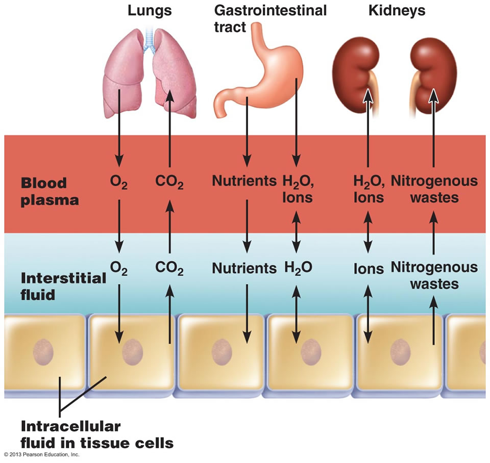
Exchange of gases, nutrients, water, and wastes between the three fluid compartments of the body.
In the image above, the ECF compartment is divisible in two compartments: (1) Plasma, the fluid portion of blood, and (2) interstitial fluid (IF), the fluid in the spaces between tissue cells.

Composition of body fluids

Electrolytes and Nonelectrolytes
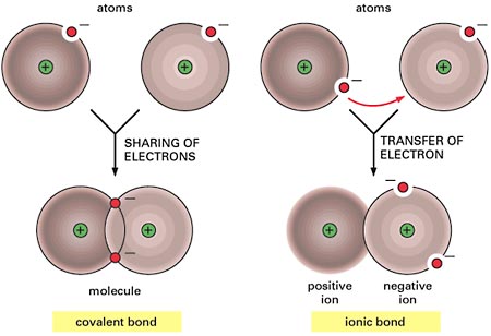
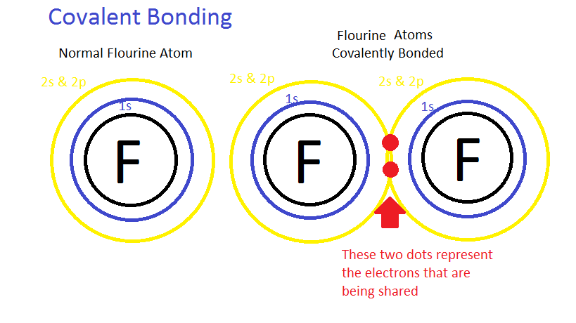
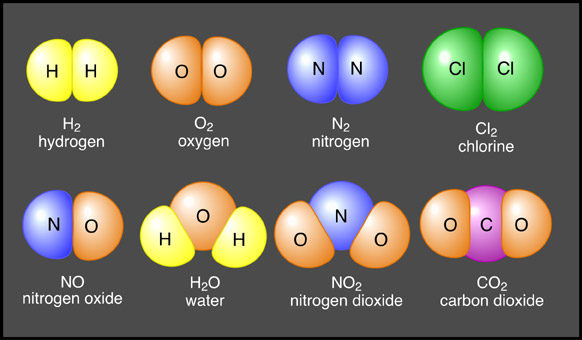
Nonelectrolytes have bonds (usually covalent bonds) that prevent them from disassociating in a solution. Because of this, no electrically charged species are created when nonelectrolytes dissolve in water. Most nonelectrolytes are organic molecules — lipids, glucose, urea, creatinine, for example.
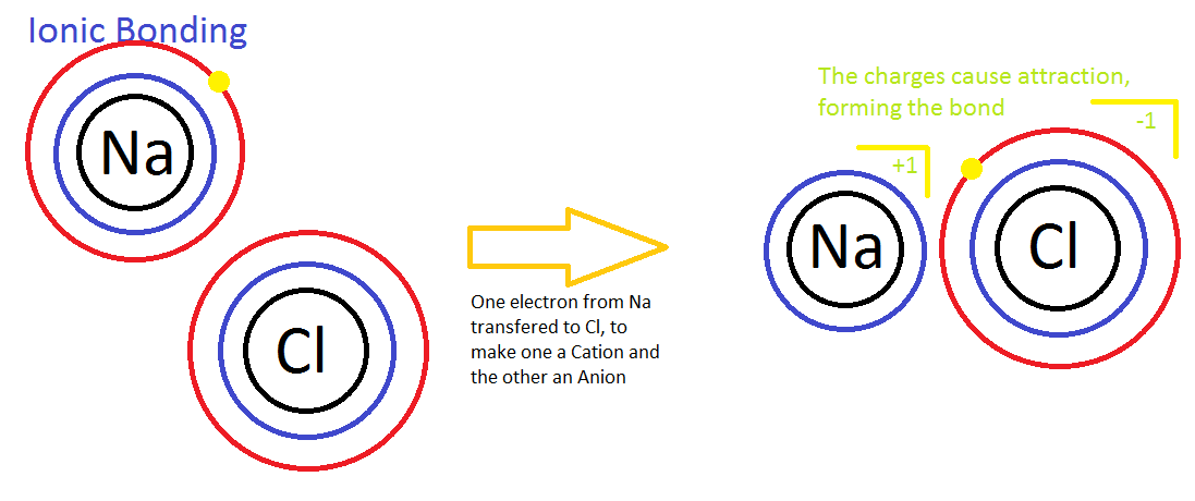
In contrast, electrolytes are chemical compounds that do disassociate into ions in water. Since ions are charged particles, they can conduct an electrical current — that’s why they’re called electrolytes! For the most part, electrolytes include organic salts, some proteins, and both organic and inorganic acids and bases.
Electrolytes have much greater osmotic power than nonelectrolytes because each electrolyte molecule disassociates into at least two ions. For instance, a molecule of sodium chloride (NaCl) contributes twice as many solute particles as glucose, and a molecule of magnesium chloride (MgCl2) contributes three times as many.
Regardless of the type of solute particle, water always moves according to osmotic gradients — from an area of lesser osmolarity to an area of greater osmolarity. For this reason, electrolytes have the greatest ability to cause fluid shifts.
fluid compartments of the human body
The major fluid compartments of the body
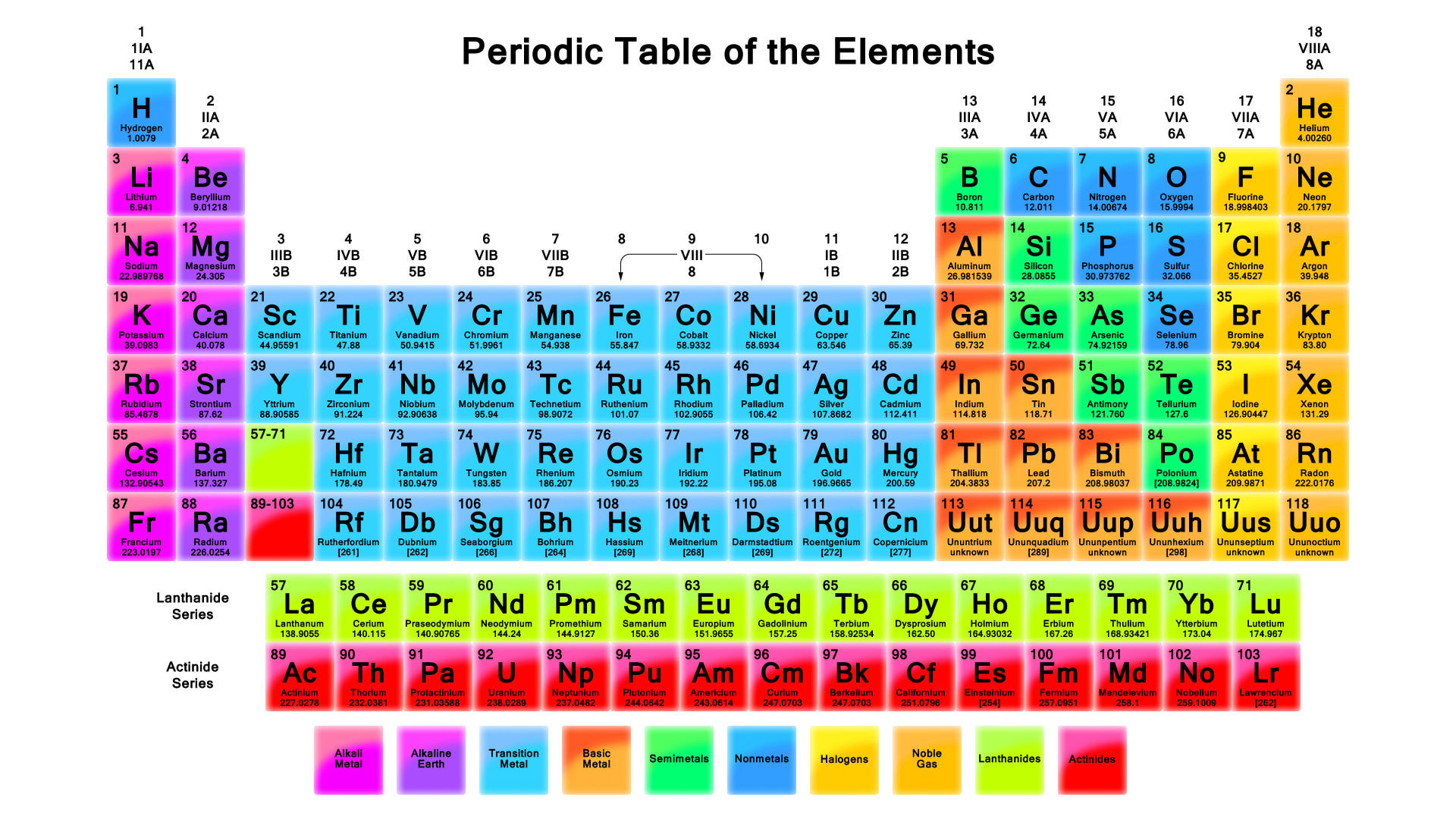
Electrolyte concentrations of body fluids are usually expressed in milliequivalents per liter (mEq/L), a measure of the number of electrical charges in one liter of solution. We can compute the concentration of any solution using the following equation: mEq/L = ion concentration (mg/L) divided by the atomic weight of the ion (mg/mmol) X the number of electrical charges on the ion. For instance to calculate the mEq/L of sodium we would determine the normal concentration of the ion in plasma, look up its atomic weight in the periodic table and plug the values into the equation: Na+ (sodium) = 3300 mg/L divided by 23 mg/mmol X 1 = 143 mEq/L. We could do the same thing for calcium: Ca2+ = 100 mg/L divided by 40 mg/mmol X 2 = 5 mEq/L.

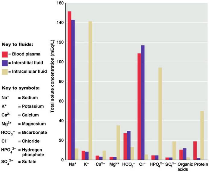
Comparison of Extracellular and Intracellular Fluids
If you look at the bar graph above you can see that each fluid compartment has a distinctive pattern of electrolytes. Beside the relatively high protein content in plasma, the extracellular fluids are very similar. The chief cation is sodium and the major anion is chloride. However, plasma contains fewer chloride molecules than interstitial fluid, because non-penetrating protein molecules are usually anions and plasma is electrically neutral. In contrast to extracellular fluid, intracellular fluid contains only small amounts of sodium and chloride. It’s most abundant cation is potassium, and its major anion is hydrogen phosphate. In the graph above, notice that sodium and potassium ion concentrations in ECF and ICF are nearly opposite. The distribution of these ions on the two sides of cellular membranes reflects the activity of cellular ATP-dependant sodium-potassium pumps, which keep intracellular sodium concentrations low and potassium concentrations high. Renal mechanisms can enforce ion distribution by secreting potassium into the filtrate as sodium is reabsorbed from the filtrate.
Electrolytes are the most abundant solutes in body fluids and determine most of their chemical and physical reactions, but they do not constitute the bulk of dissolved solutes in these fluids. Proteins and nonelectrolytes (phospholipids, cholesterol, and triglyceride) found in the ECF are large molecules. They account for around 90% of the mass of dissolved solutes in plasma and 60% in the IF, and 97% in the ICF.

Fluid Movement Among Compartments

Osmotic and Hydrostatic pressures regulate the continuous exchange and mixing of body fluids. Although water moves freely between the compartments along osmotic gradients, solutes are unequally distributed because of their size, electrical charge, or dependence on transport proteins. The image at the top of this article summarizes the exchanges of gases, solutes, and water between the three fluid compartments within the body. In general, substances must pass through both the plasma and interstitial fluid to reach the intracellular fluid. In the lungs, gastrointestinal tract, and kidneys, exchanges between the outside world and the plasma o
electrolyte composition
Electrolyte composition of blood plasma, interstitial fluid, and intracellular fluid.
ccur continuously. These exchanges alter plasma composition and volume, with plasma serving as the “highway” for delivering substances throughout the body. Compensating adjustments between the plasma and the other two fluid compartments follow quickly so that balance is restored.
Let’s review the movement of water and solutes across the boundaries between these compartments:
Exchanges between plasma and interstitial fluid occur across capillary walls. The hydrostatic pressure of blood forces nearly protein-free plasma out of the blood into the interstitial space. The filtered fluid is then almost completely reabsorbed into the bloodstream in response to the colloid osmotic pressure of plasma proteins. Under normal circumstances, lymphatic vessels pick up the small net leakage that remains behind in the interstitial space and return it to the blood.

Exchanges between the interstitial fluid and intracellular fluid occur across plasma membranes. Exchanges across the plasma membrane depend on its permeability properties. As a general rule, two-way osmotic flow of water is substantial. But ion fluxes are restricted and, in most cases, ions move selectively, by active transport or through channels. Movements of nutrients, respiratory gases, and wastes are typically unidirectional (both ways). For instance, glucose and oxygen move into the cells and metabolic wastes move out.
Many factors can change ECF and ICF volumes. Because water moves freely between compartments, however, the osmolarities of all body fluids are equal. Increasing the ECF solute content (mainly sodium chloride) causes osmotic and volume changes in the ICF — generally, a shift of water out of cells. Conversely, decreasing ECF osmolarity causes water to move into the cells. Thus, ECF solute concentration determines ICF volume.

Chemical Bonds


Chemical Bonds

When atoms combine with other atoms they are held together by chemical bonds(bonds formed between electrons of reacting atoms). These electrons occupy regions of space orbiting the nucleus called electron shells. Each electron contains one or more orbitals and each electron shell represents a different energy level (a general understanding of chemical bonds is necessary in anatomy and physiology).
A few key points to remember about electrons, orbitals, and electron shells include:
  1. Electrons furthest from the nucleus have the greatest potential energy.
  2. Electrons furthest from the nucleus are most likely to interact with other atoms.
  3. The only electrons important in chemical bonding are those in the atoms outermost energy level.

    covalent and ionic bond

The term valence shell indicates the atoms outermost electron shell or energy level (important).
When the valence shell is full (or has eight electrons), the atom is considered stable and non-reactive. If the outer shell is not full, electrons will compensate for it by gaining, losing, or sharing electrons with other atoms to achieve stability. This is when chemical bonds are formed.


Types of chemical bonds

There are three major types of chemical bonds: ioniccovalent, and hydrogen bonds.
Ionic bonding
Ionic bond– is a chemical bond between atoms formed by the transfer of one or more electrons from one atom to the other. The atom that gains an electrons is called the electron acceptor. Because electrons are electronegative, it acquires a net negative charge and is called an anion. The atom that loses electrons is called the electron donor. It acquires a net positive charge (because it loses electronegative electrons) and is called a cation. Anions and cations are formed whenever electron transfers between atoms occur. A good example of an ionic bond is table salt (NaCl). View the image above for a visual description.
covalent bonding
Covalent bond– Electrons do not have to be completely transferred for atoms to achieve stability. Instead, they can be shared so each atom is able to fill it’s valence shell. When electrons occupy a single orbital shared by both atoms, it’s called a covalent bond. For instance, in the image above two fluorine atoms are sharing a pair of electrons on their valence shell.  They are covalently bonded. Typically there are single, double, and triple covalent bonds (atoms share one, two, or three pairs of electrons).

Polar and non-polar molecules
In the covalent bonds we have discussed, the shared electrons are shared equally between the atoms, but this is not always the case. Below is a brief list of terms describing how covalent bonds may differ.
Non-polar molecules– share electrons equally and are electrically balanced (do not have separate + and – poles of charge). Carbon dioxide molecules are non-polar.
Polar molecules– Unequally share electrons and have two poles of opposite charge. Water is a good example of a polar molecule, it has a slightly negative oxygen end bonded to a slightly positive hydrogen end.
hydrogen bonds in water

Hydrogen bonds– are the weakest bond of the three types. They are more like attractions than true bonds. They occur when a hydrogen atom, already covalently linked to an electronegative atom, is attracted by an atom looking for electrons (forming a bridge between them). Hydrogen bonding is responsible for the tenancy of water molecules to cling together and form films (referred to as surface tension). This is why water beads up into spheres when sitting on a flat surface. The best example of a hydrogen bond is the bonds formed between water molecules (refer to image above).

Molecules and Compounds


Molecules and Compounds

In a recent article we discussed the importance of atoms and elements, but most atoms do not exist in a free state. Instead, they combine with other atoms through chemical bonds forming molecules and compounds.
molecule is a group of two or more atoms from the same element held together by a chemical bond. For instance, when two oxygen atoms combine, a molecule of oxygen gas forms.
When two (or more) different kinds of atoms combine, they form a compound. Compounds differ from molecules because they always contain atoms of at least two different elements. Since oxygen gas only contains one element, (oxygen) it’s considered a molecule. In contrast, water (H2O) is a compound because it contains two elements, hydrogen and oxygen. Properties of compounds are usually very different from the atoms they contain, this is an important concept to understand. For instance, water is very different from the atoms it contains (hydrogen and oxygen).


Mixtures

molecules and compoundsMixtures are substances composed of two or more components (physically mixed together). Most matter in nature exists in the form of mixtures. There are three main types of mixtures: solutions, colloids, and suspensions.

  • Solutions– are homogeneous mixtures of components (either gases, liquids, or solids). Homogeneous means the mixture has the same composition throughout.  For instance, if samples are taken from the mixture, they will have the same makeup and composition. Air and seawater are examples of homogeneous mixtures. The substance present in the greatest amount is called a solvent and is usually a liquid. Substances present in smaller amounts are called solutes. Solutes are usually dissolved by the solvent. Water is the human body’s main solvent. Most solutions in the body are true solutions (solutions containing gases, liquids, or solids dissolved in water). Saline solution is an example of a true solution.

  • Concentration of solutions– True solutions are described in terms of their concentration which can be indicated in several ways. Solutions in most colleges and hospitals are described by percentage (parts per 100). When using this method, the percentage is always based on the solute and the solvent is usually water. For instance, if the solution is 100 ml and the solute is 8 ml, the solution percentage would be 8.

  • Colloids (emulsions)are heterogeneous mixtures (their composition differs in different areas of the mixture). Colloids have several unique properties, including the ability to undergo sol-gel transformations (to change reversibly from a fluid state to a solid gel). Jell-O is an example of a colloid that changes from a liquid to a gel when refrigerated. Sol-gel transformations underlie many important cell functions including cell division.
  • Suspensions– are heterogeneous mixtures with large, visible solutes that tend to settle. An example of a suspension is a mixture of sand water (the sand settles to the bottom and completely separates until stirred again).

Distinguishing mixtures from compounds


Mixtures differ from compounds in several important ways:
  1. No chemical bonding occurs between the components of a mixture (chemical bonding does occur between compounds).
  2. Some mixtures can be separated by physical means (straining, filtering). Compounds can only be separated by chemical means (breaking bonds).
  3. All compounds are homogeneous. In contrast, some mixtures are homogeneous, while others are heterogeneous.

Atoms and Elements


Atoms and elements

All matter is composed of elements. An element is a structure that cannot be broken down into simpler substances (by ordinary methods). Some well known elements include oxygen, carbon, and iron. Currently, science recognizes 118 elements, 92 occur in nature and the other 26 are made artificially. 96% of the human body is made up of carbon, oxygen, hydrogen, and nitrogen. These are known as the four main elements of the human body. 20 other elements are also present in the body, some in trace amounts. The periodic table lists all known elements and explains the properties of each.
All elements are composed of smaller particles called atoms. Every elements atoms differ from those of other elements giving it unique physical and chemical properties.
  • Physical properties– are properties we can detect with our senses (touch, smell) or measure (boiling point).
  • Chemical properties– is how atoms interact with each other (bonding behavior).
Each element is designated with an atomic symbol. An atomic symbol is a one or two letter (usually the first letter/s of an elements name) shortened name. For instance, O stands for oxygen, C stands for carbon.


Atomic structure

Periodic Table

Atoms are small, but atoms are made up of even smaller particles. These subatomic particles differ in mass, electrical charge and position. Below is a description of each.
Nucleus- each atom has a central nucleus. The central nucleus contains protons and neutrons bound together. The nucleus is surrounded by electrons (orbiting).
  1. Proton– is a particle with a positive electrical charge.
  2. Neutron– is a particle with a neutral charge. Because the nucleus is made up of protons and neutrons, it has an overall positive charge.
  3. Electron– is a particle with a negative charge. Electrons are extremely small (1/2000 the mass of a proton).
All atoms are electrically neutral because the number of protons (+) is balanced out by the number of electrons (-). For example, iron has 26 protons and 26 electrons (making its electrical charge neutral). The number of electrons and protons is always equal in an atom.

Common elements in the human body

  • Oxygen (O)- A major component of organic (carbon-containing) and inorganic (non-carbon containing) molecules. Oxygen is needed for the production of ATP during cellular respiration.
  • Carbon (C)– Component of organic molecules (carbs, fats, proteins, molecules).
  • Hydrogen (H)– Component of organic molecules. Influences pH of body fluids.
  • Nitrogen (N)– A component of proteins and nucleic acids.
  • Calcium (Ca)– found as salt in bones and teeth.
  • Phosphorus (P)– Part of calcium phosphate salts in bones and teeth.
  • Potassium (K)– Necessary for conduction of nerve impulses and muscle contraction. Major positive ion in cells
  • Sulfur (S)– Component of proteins (especially muscle proteins).
  • Sodium (Na)– Important in water balance, muscle contractions, and nerve impulses. Most abundant positive ion in extracellular fluid.
  • Chlorine (Cl)– Most abundant negative ion in extracellular fluid.
  • Magnesium (Mg)– Important in several metabolic reactions. Present in bone.
  • Iodine (I)– Used to make functional thyroid hormones.
  • Iron (Fe)– Component of hemoglobin (transports oxygen) and some enzymes.
  • There are trace elements in the body also. Trace elements are elements required in small (minute) amounts. A few examples include cobalt (Cr), flourine (F), copper (Cu), silicon (Se) and more.

    Planetary model of the atom
    orbital model atom
    The planetary model of an atom illustrated above depicts electrons moving around the central nucleus in a fixed circular orbit. But even though the orbits are fixed, the exact location of electrons isn’t known because they follow unknown trajectories around the nucleus. An orbital is defined as regions around the nucleus electrons are likely to be found most of the time. Using the orbital model is a useful tool in predicting the chemical behavior of atoms.
    Atoms of different elements are composed of different numbers of protons, neutrons, and electrons. For instance, hydrogen has one proton, one electron, and no neutrons. Helium has two protons, two neutrons, and two electrons and the list goes on. So, how do we recognize and label these differences? Science uses atomic number, atomic weight, and mass number to do this. If you take a look at the periodic table, you can locate all of them.
    • Atomic number– is equal to the number of protons int he nucleus of an atom. It is written as a subscript to the left of the atomic symbol. For instance, hydrogen’s atomic number would be 1 because it has one proton.
    • Mass number– is the sum of the atoms protons and neutrons. For example, helium has two protons and two neutrons so its mass number is 4.
      Helium atomic mass and number
    Isotopes– are structural variations of an element. Almost all known elements have two or more structural variations (isotopes). Isotopes have the same number of protons and electrons but differ in the number of neutrons they contain. Earlier, we said hydrogen has a mass number of 1 (which it does) but this is not always the case. Hydrogen has several isotopes, it’s most abundant contains a mass number of 1, but others contain mass numbers of 2 and 3.
    Atomic weight– is an average of the relative weights (mass numbers) of all the isotopes of an element. In most cases, the atomic weight is equal to the mass number of its most abundant isotope.

    Radioisotopes-the process of atomic decay is called radioactivity. This happens when an atom is unstable and decays spontaneously into a more stable form. Isotopes that cause radioactivity are called radioisotopes. Radioisotopes are valuable tools used in biology and medicine. For example, Iodine 131 is used to determine the size and activity of the thyroid gland. It’s also used to detect thyroid cancer, but radioisotopes can harm the body too. For instance, inhaled particles from decaying radon can cause lung cancer.

The Tissue

Medical Yukti

Medical Yukti

Blood


Vestigial-organs

Respiratory-system

Nutrition-and-Health

PCOS

Vulvar-Cancer

Header Ads Widget